Session Abstract – PMWC 2023 Silicon Valley
Track Chair: Sean Khozin, CancerLinQ
- PMWC Award Ceremony:
Pioneer Honoree: Daniela Ushizima, LBNL, UCSF, UCB & Lea Grinberg, UCSF
- Pattern Recognition and Content Quantification in Early-stage Disease Diagnosis (PANEL)
Chair: Daniela Ushizima, LBNL, UCSF, UCB
- Lea Grinberg, UCSF
- Suzanne Baker, LBL - Second Phase of AI Imaging is Clinical Validation
- Sean Khozin, CancerLinQ
- Large-Scale Spatial Omics Imaging Analysis To Decipher The Tumor Microenvironment
- Joseph Lehar, Owkin - Latest Achievement in AI Imaging Technology in the area of Radiology and Pathology
Chair: Lars Coster, Icometrix
- Joachim H. Schmid, NanoString Technologies
- Fedaa Najdawi, PathAI
- Nurit Paz Yaacov, Imagene
- Neil Weisenfeld, 10x - AI in Radiology & Pathology Applications (Clinical Research)
Chair: Mirabela Rusu, Stanford
- Saeed Hassanpour, Dartmouth
- Jeanne Shen, Stanford
- Pratik Mukherjee, UCSF - PMWC NCI Showcase
- Omid Moghadam, Rapid - PMWC Showcase
- Amanda Hanson, Spartanburg Regional Healthcare System
Session Chair Profile
Biography
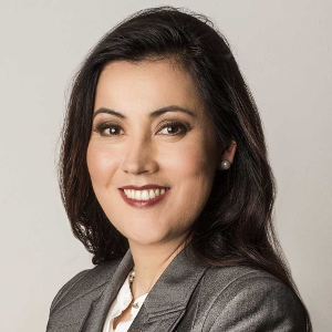
Dani Ushizima is a Computer Scientist focused on Computer Vision and Machine Learning algorithms to characterize materials toward self-driving labs. Her research has impacted projects that depend on experimental data coming from instruments reliant on x-ray, electron, confocal, and other light-matter interactions. In 2015, Ushizima received the U.S. DOE Early Career Research award to work on pattern recognition for scientific images. In 2021, she was honored by 3M as one of the top "25 Latina in Science" for biomedical research. Also in 2021, Ushizima and colleagues received the Berkeley Lab Halbach award for creating machine-learning-based techniques in support to the Advanced Light Source. Currently, Ushizima leads research projects in the Center for Advanced Mathematics for Energy Related Applications (CAMERA). Jointly with UCSF Grinbergs lab, she has developed a new technique for detecting biomarkers of Alzheimer’s disease and for supporting measurement of the efficacy in experimental treatments.
Session Chair Profile
Biography
Dr. Lars Costers is product manager at icometrix, a Belgian scale-up company founded in 2011 and leader in the field of imaging AI. Lars is a neuroscientist with a PhD in medical sciences, responsible for building digital health tools and platforms that enable the implementation of precision medicine in the field of neurology.
Icometrix offers a portfolio of AI solutions to assist healthcare with various challenges in neurological disorders such as multiple sclerosis, brain trauma, epilepsy, stroke, dementia, and Alzheimer's disease. From deep-learning-based image quantification to mobile apps that allow the telemonitoring of patients using cognitive assessments, PROs and passive monitoring.
We have produced 9 FDA/CE approved solutions, 3 approved patents on core technology, over 230 peer-reviewed articles, and are integrated with over 200 clinical sites.
Talk
Time is Brain Precision Medicine in Neurology
The societal and economic burden of chronic neurological conditions, such as Alzheimers disease and Multiple Sclerosis, is immense. However, current care pathways are variable and disease monitoring is qualitative and subjective. Hence, there is a huge need for digital health solutions and AI to enter the era of precision medicine in neurology.
Session Chair Profile
Biography
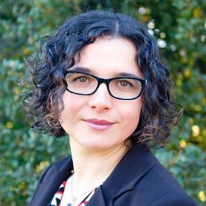
Dr. Mirabela Rusu received a Master of Engineering from the National Institute of Applied Sciences, France and her MS and PhD in Computational Biomedicine from University of Texas Houston. During her postdoctoral training at Case Western Reserve University, Dr. Rusu developed computational tools for the integration and interpretation of multi-modal medical imaging data. Dr. Rusu joined GE Global Research in 2015 as an Image Analysis Scientist and developed analytic methods for biomedical data interpretation. Since 2018, Dr. Rusu leads the Personalized Integrative Medicine Laboratory (http://pimed.stanford.edu) which focuses on developing analytic methods to spatially align radiology and pathology images in patients that undergo surgery for cancer treatment. Moreover, her team uses the registered data to label the radiology images and extract pathomic MRI biomarkers that allow the training of deep learning models to automatically detect and distinguish indolent from aggressive prostate cancer on MRI.
Talk
Bridging the gap between radiology and pathology images
Speaker Profile
Biography
The last 5 years Amanda’s primary focus at Spartanburg Regional has been to pioneer Precision Medicine and implement Precision Pathology to ensure the right treatment for the right patient at the right time. She spent over a decade as a Clinical Molecular Biology Scientist performing high complexity molecular testing before taking on her leadership role at a community-based hospital. Here she has worked extensively with Pathologists, Oncologists and Technologists using her unique background and passion for personized medicine to ensure the highest quality of care for her local rural community.
Talk
Developing a Precision Medicine Program in a Community Hospital
In this presentation I will be discussing how Spartanburg Regional launched a Precision Medicine Program in under 5 years that among many beneficial things has resulted in our Oncologists receiving their actionable variant sequencing results over a week faster than the national average.
Speaker Profile
Biography
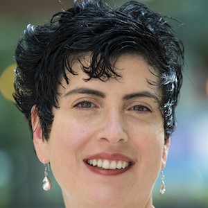
Dr. Lea Tenenholz Grinberg is a neuropathologist specializing in brain aging, most notably, Alzheimer’s diseases. She is a Full Professor and a John Douglas French Alzheimer’s Foundation Endowed Professor at the UCSF Memory and Aging Center, where she co-led the Neuropathology Core. She is also a Professor of Pathology at the University of Sao Paulo. Dr. Grinberg is the recipient of several awards and have co-authored over 260 scientific papers. The Grinberg Lab investigates factors influencing clinical expression of Alzheimer’s pathology and other tauopathies to lead to better diagnostic tools and therapeutic targets that minimize clinical decline in AD by following up on Dr. Grinberg’s initial discoveries of brainstem vulnerability in Alzheimer’s disease. Her discoveries had changed the understanding on the basis of sleep disturbances in these diseases. The Lab combines classical quantitative neuropathological techniques with advanced computer vision tools and multiplex molecular probing in postmortem human tissue
Talk
Unveiling the Basis of Neuroimaging Signal with Histology
neuroimaging findings is often based on assumptions because of its lower resolution compared to microscopy. We developed a computational and histological pipeline to process and stain whole human brain volumes, 3D reconstruct the histological volume at microscopical level and perfectly align histology-based cytoarchitectonic and protein deposition 3D maps to their neuroimaging counterparts.
Speaker Profile
Biography
Over 20 years experience as an innovator and executive, focused on using data and digital technologies to transform health care. Before my current activities at Owkin, venture and board member/advisor to multiple organizations, I led cross-functional teams and drove scientific projects at J&J/Janssen (as VP of Data Science), Google/Verily, Novartis, and CombinatoRx.
Talk
Data science and precision medicine at Owkin
I will present an overview of biomedical needs addressable by data science, and how Owkin is advancing precision medicine. I will focus on capabilities and promising projects we are undertaking, including Owkins digital spatial profiling initiative in collaboration with multiple academic institutes.
Speaker Profile
Biography
Dani Ushizima is a Computer Scientist focused on Computer Vision and Machine Learning algorithms to characterize materials toward self-driving labs. Her research has impacted projects that depend on experimental data coming from instruments reliant on x-ray, electron, confocal, and other light-matter interactions. In 2015, Ushizima received the U.S. DOE Early Career Research award to work on pattern recognition for scientific images. In 2021, she was honored by 3M as one of the top "25 Latina in Science" for biomedical research. Also in 2021, Ushizima and colleagues received the Berkeley Lab Halbach award for creating machine-learning-based techniques in support to the Advanced Light Source. Currently, Ushizima leads research projects in the Center for Advanced Mathematics for Energy Related Applications (CAMERA). Jointly with UCSF Grinbergs lab, she has developed a new technique for detecting biomarkers of Alzheimer’s disease and for supporting measurement of the efficacy in experimental treatments.
Speaker Profile
Biography
Dr. Lea Tenenholz Grinberg is a neuropathologist specializing in brain aging, most notably, Alzheimer’s diseases. She is a Full Professor and a John Douglas French Alzheimer’s Foundation Endowed Professor at the UCSF Memory and Aging Center, where she co-led the Neuropathology Core. She is also a Professor of Pathology at the University of Sao Paulo. Dr. Grinberg is the recipient of several awards and have co-authored over 260 scientific papers. The Grinberg Lab investigates factors influencing clinical expression of Alzheimer’s pathology and other tauopathies to lead to better diagnostic tools and therapeutic targets that minimize clinical decline in AD by following up on Dr. Grinberg’s initial discoveries of brainstem vulnerability in Alzheimer’s disease. Her discoveries had changed the understanding on the basis of sleep disturbances in these diseases. The Lab combines classical quantitative neuropathological techniques with advanced computer vision tools and multiplex molecular probing in postmortem human tissue
Speaker Profile
Biography
Suzanne Baker got her bachelor degree in Biomedical engineering from Tulane University and her PhD in Vision Science from University of California, Berkeley. Her research focuses on methods development in brain positron emission tomography (PET) in aging and dementia. Her primary interests are tracer evaluation, pharmacokinetic modeling, harmonizing multi-site data, reference region analysis, between tracer comparison and partial volume correction. She serves as the PET core for many multi-center studies. She has performed preliminary evaluation of Flortaucipir and JNJ067 tau PET tracers. She consults for Genentech on their tau PET tracer. She recently was awarded a $40 million NIH grant to compare Flortaucipir and MK6240 tau PET tracers in a head-to-head cohort, which will include comparisons to measurements of tau in plasma.
Talk
Considerations in methods for tau PET quantification
A useful PET tracer has high affinity for the protein of interest and has none-to-low binding to anything else. In aging and dementia, the correlation of tau accumulation with cognitive decline makes it an interesting target for therapies. Here Ill explore methods of amplifying the signal and decreasing the noise.
Speaker Profile
Biography
Sean Khozin is an oncologist and data scientist. He is a globally recognized leader in building solutions at the intersection of biology, technology, and AI/machine learning to advance biomedical research and therapeutic development.
Talk
Artificial Intelligence as a Tool to Reclassify Diseases
Despite the recent advances in the development of new medicines, many patients do not derive any benefit from available therapies and diseases like metastatic cancers remain largely incurable. Catalyzing the next wave of biomedical breakthroughs and precision therapies requires a paradigm shift in classification of diseases using AIML solutions.
Speaker Profile
Biography
Joachim Schmid recently joined NanoString as Vice President of Spatial Informatics & AI and oversees the development of innovative software solutions and data analysis methods for spatial biology. Previously he worked in various R&D leadership positions at Tripath Imaging, Dako, and Roche Tissue Diagnostics.
Speaker Profile
Biography
Dr. Najdawi is a board certified anatomical pathologist specialized in gastrointestinal and endocrine pathology. She works at the intersection of Artificial Intelligence and Anatomical Pathology at PathAI, where she is using her pathology expertise to guide a multidisciplinary team with a mission to improve patient outcomes with AI-powered pathology.
Dr. Najdawis pathology journey started in Australia and she completed her AP training and two fellowships in gastrointestinal and endocrine pathology at Brigham and Women’s Hospital, Harvard Medical School. She has over 12 years of experience in the medical field, and is certified to practice medicine in the USA, Australia, and Jordan. She has multiple peer-reviewed publications and presented her work at national and international meetings. Her current research is focused on leveraging AI and digital pathology tools to empower precision pathology.
Speaker Profile
Biography
Neil leads analysis software pipeline development for the Visium line of spatial transcriptomics products at 10x. He has over 25 years experience in image analysis algorithm development for diverse applications including functional brain imaging, image-guided neurosurgery, neonatal brain MRI, and pathology image analysis. Prior to joining 10x, he was a senior computational biologist at the Broad Institute working on algorithms for de novo genome assembly.
Talk
High-resolution mapping of the breast cancer tumor micro environment
We describe new methods that bring together single-cell gene expression (Chromium Fixed RNA Profiling), spatial transcriptomics (Visium CytAssist), and automated, microscopy-based in situ technology (Xenium In Situ) to better characterize the breast cancer tumor microenvironment leading to new biological insights about the trajectory of disease.
Speaker Profile
Biography
Dr. Hassanpour’s research is focused on building novel machine learning and multimodal data analysis methods to inform precision health. His lab has been a pioneer in advancing digital pathology through deep learning methodologies. He has led multiple NIH-funded research projects on developing new machine learning models for medical image analysis and clinical text mining to improve diagnosis, prognosis, and personalized therapies. His research has resulted in numerous publications, software, and datasets that are widely recognized and received multiple awards, including the 2019 Agilent Early Career Professor Award for breakthroughs in digital pathology. Before joining Dartmouth, he worked as a Research Engineer at Microsoft. Dr. Hassanpour received his Ph.D. in Electrical Engineering with a minor in Biomedical Informatics from Stanford University and completed his postdoctoral training at Stanford Center for Artificial Intelligence in Medicine & Imaging.
Talk
AI for Digital Pathology
With the recent expansions of whole-slide digital scanning, archiving, and high-throughput tissue banks, digital pathology is primed to benefit dramatically from AI. This talk will cover several clinical applications of AI for characterizing histopathological patterns on high-resolution microscopy images for the classification, prognosis, and treatment of cancerous and precancerous lesions.
Speaker Profile
Biography
Dr. Mukherjee is a board-certified clinical neuroradiologist whose research has centered on technical development and basic and clinical neuroscience applications of methods for mapping tissue microstructure, connectivity and function in the human brain. Twenty years ago, he led some of the initial work characterizing neonatal, infant and childhood brain development using diffusion tensor imaging (DTI). Recent work includes the application of advanced diffusion MRI, resting state fMRI and MEG to study cerebral connectivity, including the whole-brain network known as the “connectome”, in human brain development, neurodevelopmental disorders and traumatic brain injury (TBI). He has special experience in standardizing structural MRI, dMRI, and fMRI pulse sequences and scan protocols for large-scale multi-site projects and managing the resulting Big Data using cutting-edge informatics platforms and machine learning analytics, which is essential for the clinical translation of new imaging technology.
Talk
AI in Human Brain Imaging
Speaker Profile
Biography
Dr. Nurit Paz-Yaacov is the Chief Scientific Officer at Imagene AI - a BioMed startup company with groundbreaking technology in the field of precision oncology using Artificial Intelligence.Dr. Paz-Yaacov brings more than 20 years of multidisciplinary scientific research in human genetics and genomic sequencing and interpretation, with an emphasis on cancer research.Prior to her role at Imagene AI, Dr. Paz-Yaacov was the Head of Scientific Research at Genoox, a cloud platform that provides actionable interpretation for real-time/real-life genomic data.Dr. Paz-Yaacov holds B.Sc, M.Sc, and Ph.D. degrees from Tel-Aviv University and completed her Postdoctoral studies at Bar-Ilan University. Her studies, and research in the fields of cancer, applied genomics, and epi-genomics, received recognition through numerous publications and prizes.
Speaker Profile
Biography
Dr. Shen is a board-certified pathologist and Associate Director of Pathology for the Stanford Center for Artificial Intelligence in Medicine and Imaging. She has served as a principal investigator and collaborator on multiple academic and industry-sponsored studies in digital and computational pathology and oncology, and as a medicolegal, startup, and venture capital consultant focused on applications in digital pathology and AI. She received her BS in Biological Sciences from Stanford University and MD from Washington University in St. Louis, and completed Anatomic and Clinical Pathology (AP/CP) and fellowship training in Gastrointestinal and Liver Pathology at Brigham and Women’s Hospital, Harvard Medical School, along with additional training in biodesign innovation as a Biodesign Faculty Fellow at Stanford University (http://profiles.stanford.edu/jeanne-shen)
Talk
AI-Based Tumor-Infiltrating Lymphocyte Assessment as a Practical Immuno-Oncology Biomarker
Speaker Profile
Biography
Omid Moghadam is the founder and CEO of Namida Lab, a revenue-stage diagnostics company specializing in developing and commercializing liquid biopsy tests for early cancer detection. Namidas first product, Auria, is a direct-to-consumer proteomic test allowing women to take charge of their breast health.
Hes an inventor, entrepreneur, venture investor, and educator. He specializes in launching new ventures with social impact in health and technology. Moghadam has inventions in medical imaging, cryptography, microprocessor design, medical devices, diagnostics, digital photography, data-science and communications.
Hes the past founder or co-founder of nine companies in Healthcare IT, genomics, diagnostics, and medical imaging, and has held executive positions at Intel, Eastman Kodak, and CTG-AMS Corporations.
He formerly served on the advisory boards of Robert Wood Johnson Foundation, Childrens Hospital Boston, Abbott Corporation, and California Healthcare Foundation and has held academic positions at Harvard Medical School department of biomedical informatics, as well as an Executive-In-Residence position at UCLA.
Speaker Profile
Biography
Dr. Lea Tenenholz Grinberg is a neuropathologist specializing in brain aging, most notably, Alzheimers diseases. She is a Full Professor and a John Douglas French Alzheimers Foundation Endowed Professor at the UCSF Memory and Aging Center, where she coled the Neuropathology Core. She is also a Professor of Pathology at the University of Sao Paulo. Dr. Grinberg is the recipient of several awards and have coauthored over 260 scientific papers. The Grinberg Lab investigates factors influencing clinical expression of Alzheimers pathology and other tauopathies to lead to better diagnostic tools and therapeutic targets that minimize clinical decline in AD by following up on Dr. Grinbergs initial discoveries of brainstem vulnerability in Alzheimers disease. Her discoveries had changed the understanding on the basis of sleep disturbances in these diseases. The Lab combines classical quantitative neuropathological techniques with advanced computer vision tools and multiplex molecular probing in postmortem human tissue
Talk
Unveiling the Basis of Neuroimaging Signal with Histology
neuroimaging findings is often based on assumptions because of its lower resolution compared to microscopy. We developed a computational and histological pipeline to process and stain whole human brain volumes, 3D reconstruct the histological volume at microscopical level and perfectly align histology-based cytoarchitectonic and protein deposition 3D maps to their neuroimaging counterparts.

